

What Is A Sporophyte Apex
Introduction
The ability to decrease h2o loss was disquisitional for the development and survival of plants in terrestrial environments (Graham, 1993). On the aerial organs of plants, water loss is decreased by the cuticle, a modified cell wall region consisting of polysaccharides and a polymer matrix of cutins/cutans embedded with waxes and phenolics, in addition to waxes deposited on the outer surface of the matrix (Domínguez et al., 2011; Guzmán et al., 2014). The cuticle occurs on all land plants, including mosses (Busta et al., 2016), and information technology is important for protection from ultraviolet (UV) radiation (Krauss et al., 1997; Holmes and Keiller, 2002; Pfündel et al., 2006), self-cleaning of photosynthetic surfaces (Barthlott and Neinhuis, 1997), and prevention of pathogen attacks (Campbell et al., 1980). Cuticles occur in all lineages of land plants and play critical roles fifty-fifty in the earliest diverging lineages (i.e., liverworts, hornworts, and mosses). On the vegetative gametophytes of these plants, cuticles create water-free regions on the external plant trunk that facilitate gas exchange, which occurs direct through the epidermal cell walls, given the absenteeism of stomata (Schönherr and Ziegler, 1975; Thomas et al., 1996). The cuticle as well maintains internal hydration in mosses for both the gametophytes of endohydric taxa (Proctor, 1979) and for the relatively long-lived sporophytes (Budke et al., 2013).
Moss sporophytes begin development within the female reproductive organs (archegonia) and are protected by the surrounding leaves of the maternal gametophyte. Every bit development progresses, the sporophyte increases in height and its exposure to dehydration stress is magnified as it emerges from the leaves of the maternal gametophyte and the protective influence of the laminar boundary layer (Proctor, 1980, 1982; Rice et al., 2001; Rice and Schneider, 2004). Immature moss sporophytes take a sparse cuticle that structurally is unable to protect them from dehydration (French and Paolillo, 1975; Budke et al., 2012). Protection for the dehydration-sensitive sporophyte noon during the critical developmental stage of stalk-building is provided by the calyptra, a cap of maternal gametophyte tissue that has a thick, multilayered cuticle (Budke et al., 2011). This cuticle-covered cap forms early on in development and protects the moss sporophyte apex until the sporophyte later on develops a thicker cuticle, during sheathing maturation (Budke et al., 2012). The calyptra cuticle is functionally important for sporophyte survival, evolution, and fitness (Budke et al., 2013).
Sporophyte morphology is highly variable across the 12,500 species of mosses with sporophyte heights ranging from over 9 cm tall in Polytrichum Hedw. (Smith Merrill, 2007) to less than 1 mm in Micromitrium Austin (Goffinet et al., 2011). Sporophyte superlative can vary within a family or genus, potentially accompanying a shift to drier or seasonally moist habitats or in the other farthermost a shift to aquatic habitats (Vitt, 1981). Within the Funariaceae Schwägr., a wide range of sporophyte sizes is represented (Fife, 1982). Ranging from taxa with a tall stalk elevating the capsule loftier above the maternal plant (eastward.g., Funaria hygrometrica Hedw.) to species that substantially lack a stalk and thus have a sheathing, which at maturity is immersed among the leaves originally surrounding the female person sex organs [due east.grand., Aphanorrhegma serratum (Hook and Wilson) Sull.]. This wide diversity among closely related species makes the Funariaceae an platonic system for comparative studies of the sporophyte and its cuticle.
Growing under equivalent ecological weather condition, shorter sporophytes would exist predictable to experience lower levels of dehydration stress compared to taller sporophytes due to the closely surrounding leaves and the protection afforded by the laminar boundary layer of the adjacent substrate (Proctor, 1980, 1982; Rice et al., 2001; Rice and Schneider, 2004). Released from the selective pressures of dehydration stress, the part of the cuticle in protection of the sporophyte may become unnecessary. Thus, retention of a thick cuticle in taxa with curt sporophytes may point toward an culling functional importance, such as protection from UV radiation (Krauss et al., 1997; Holmes and Keiller, 2002; Pfündel et al., 2006). The cuticle is a costly resource investment (Poorter and Villar, 1997). Decreasing the investment in this protective layer past either the maternal plant (i.due east., the calyptra) or the offspring sporophyte frees up resources to devote to other processes that heighten fettle and thereby ultimately reproductive success, such as spore production.
In this study, nosotros explore the relationship between cuticle thickness and sporophyte acme every bit a proxy for aridity stress in taxa with sporophytes across a range of sizes. We admit that studies of tracheophyte leaves and fruits accept not confirmed Fick's first law, that states cuticular permeability should be related to the thickness of the cuticular membrane (Norris, 1974; Riederer and Schreiber, 2001). The quantities of waxes and/or cutins take also not been shown to predict cuticular permeability (Riederer and Schreiber, 2001; Yeats and Rose, 2013). Instead, the ratios between waxes classes, rather than the overall quantities, have been shown to correlate with cuticular permeability (Parsons et al., 2012). Analyzing the structural variation in bryophyte cuticles insufficiently across taxa and development is the first stride in exploring Fick's starting time police in bryophytes. Bryophytes have cuticles that are orders of magnitude thinner than vascular plants (Jeffree, 2006) and are separated by at to the lowest degree 420 million years of evolution from their nearly recent common ancestors (Clarke et al., 2011). Alternative relationships may sally for bryophytes, potentially including a correlation between cuticle thickness and permeability, which currently remains to be tested.
We know that the maternal institute invests in a relatively thick calyptra cuticle that protects young sporophytes from the stress of dehydration in F. hygrometrica (Budke et al., 2011, 2013). First, nosotros sought to ostend (i) that immature sporophytes of all species have a very thin cuticle during early development and (two) that sporophytes develop a thick cuticle afterward in their development. Given the cuticle developmental patterns in (i) and (ii) are confirmed, we hypothesize (three) that the maternal investment in the calyptra cuticle would correlate with sporophyte height: the calyptra cuticle will exist thinner for species with shorter sporophytes and thicker for species with taller sporophytes. We hypothesize that this investment in a calyptra cuticle volition occur early and persist throughout sporophyte development. These hypotheses are based on shorter sporophytes likely encountering less dehydration stress during development, compared to taller sporophytes. Additionally we hypothesize (iv) that species with shorter sporophytes will have a thinner sporophyte cuticle compared to species with taller sporophytes. To address these hypotheses, we compared the development of the cuticle on the sporophyte and calyptra for four moss species in the Funariaceae that cover a range of sporophyte and calyptra sizes.
Materials and Methods
Written report Taxa
Mosses in the Funariaceae have very like leafy gametophyte morphologies. The morphological diversity in this family lies in the maternal gametophyte calyptra and sporophyte (Fife, 1982). 4 Funariaceae species with contrasting calyptra and sporophyte morphologies were cultured in the laboratory for this written report. Aphanorrhegma serratum (NY Buck #49500) is very small with a sporophyte with a globose capsule that is less than one mm tall that remains immersed amid the leaves of the maternal gametophyte even at maturity (Figure 1F). The calyptra of A. serratum is a small cap with several lobes at the base and overall is less than one-half a centimeter long (Figures 1A,F). Physcomitrellopsis africana Wagner and Broth. ex. Dixon (CONN Goffinet #10326) is a slightly larger moss with a sporophyte ending in an elliptic capsule that is less than three mm tall, and that remains relatively immersed among the leaves of the maternal gametophyte even at maturity (Figure 1H). The calyptra of P. africana, at less than 2 mm in length, has a short rostrum at the apex and an inflated base beneath that covers a bulk of the sporophyte capsule at maturity (Figures 1B,H). Physcomitrium pyriforme (Hedw.) Hampe (CONN Goffinet #9276) has a relatively tall sporophyte (around 15 mm) with a globose-pyriforme capsule that is exerted higher up the maternal gametophyte at maturity (Figure 1J). The calyptra of P. pyriforme is 2–iii mm long, ends in a rostrum and is deeply split at the base in 2–4 lobes (Figures 1C,J). Funaria hygrometrica (CONN Budke #142) has a very tall sporophyte, which can achieve heights of 80 mm elevating the asymmetrically curved sheathing far above the maternal gametophyte at maturity (Figure 1L). The calyptra of F. hygrometrica, which tin can exist iii–5 mm long, has a long rostrum and a wide inflated base of operations that is split past a unmarried slit (Figures 1D,L).
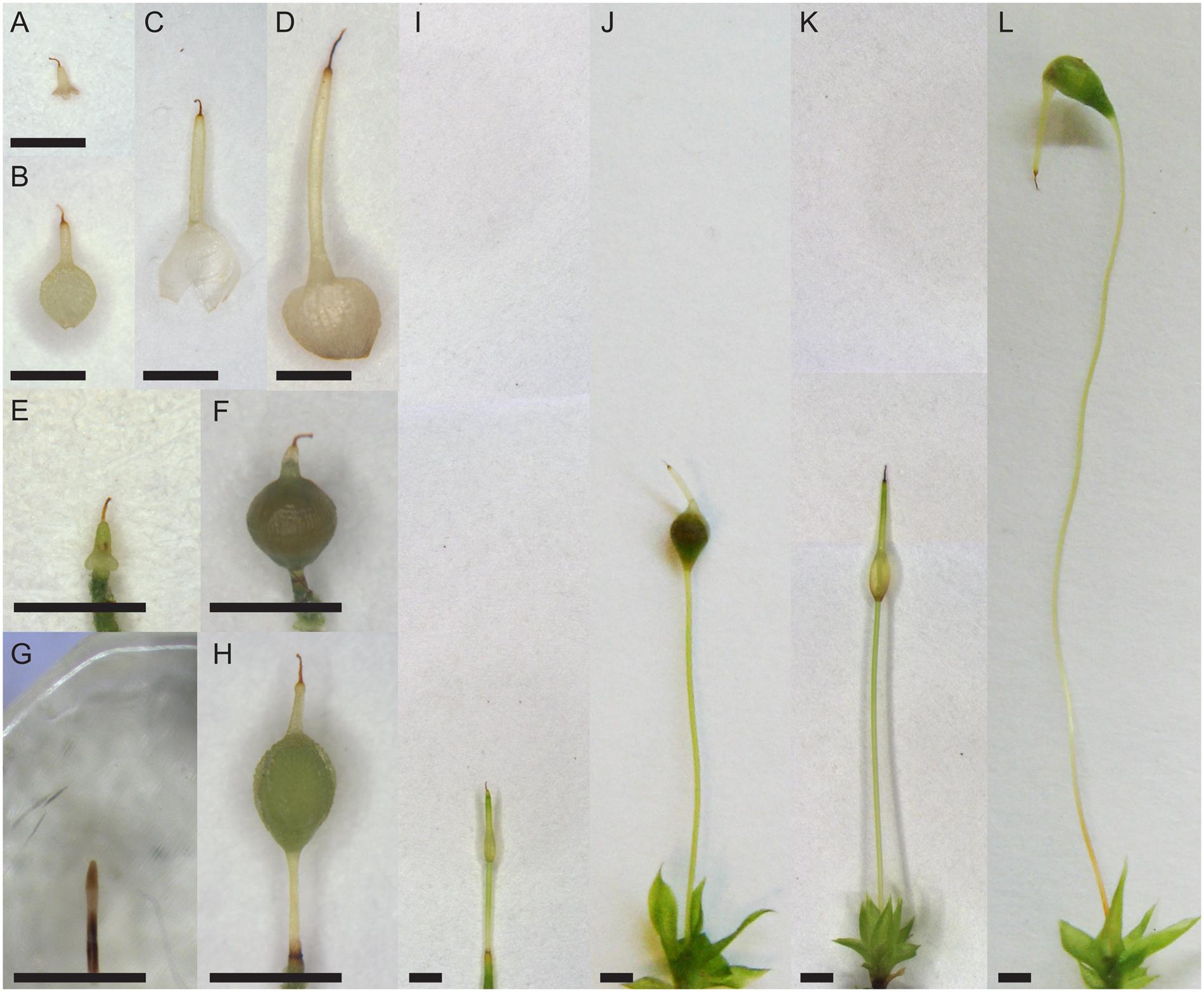
Effigy 1. Morphology of Funariaceae species. (A–D) Older calyptrae. (E,Thousand,I,K) Young sporophytes with apex covered past calyptrae, except (G) without calyptra. (F,H,J,L) Older sporophytes with calyptra on the height. (A,Eastward,F) Aphanorrhegma serratum. (B,Thousand,H) Physcomitrellopsis africana. (C,I,J) Physcomitrium pyriforme. (D,K,L) Funaria hygrometrica. Scale bars: 1 mm.
Leafy gametophytes were grown from spores of the original populations on a rich sandy loam soil mix at room temperature (approximately 22–25°C) under sixteen h of daylight in PlantCon plant tissue culture containers (MP Biomedicals, Solon, OH, Us). Aphanorrhegma serratum produced gametangia and subsequently sporophytes after ii–iii months at room temperature. The other iii taxa were grown at room temperature for 4 months, and so cold treated at 10°C with viii h daylight for 2 months to stimulate gametangial development. Deionized water was added to populations with gametangia, roofing the leafy gametophytes, for 24 h to raise fertilization. After the water was removed, plants remained in the cold growth chamber for one additional week. Populations were then placed at room temperature under sixteen h of daylight to facilitate sporophyte development. Calyptra and sporophytes from each population were harvested at ii developmental stages, young with a spear-shaped, unexpanded sporophyte, and older with an expanded sporangium containing spores, from individuals located toward the eye of the container to eliminate any potential edge effects (Figure i).
Manual Electron Microscopy
To investigate cuticle ultrastructure the sporophyte apex, including the region of the expanded sporangium, and their associated calyptra were collected. The older sporangia and calyptrae were split longitudinally to facilitate fixation and infiltration. All tissues were immediately placed into fixative (1.5% glutaraldehyde, i.5% formaldehyde in 0.05 M PIPES buffer, pH 7.0; Renzaglia et al., 1997) for iv–8 h nether ambient conditions, then overnight under vacuum, for a total of 24 h of fixation. Tissues were rinsed in 0.05 M PIPES buffer twice for 20 min each and kept in buffer overnight at four°C in the dark. Tissues were then rinsed in 0.05 K PIPES buffer in one case for 20 min. Osmium fixation (ii.0% OsO4 in 0.05 K PIPES buffer, pH seven.0) was carried out for 2 h in the nighttime followed by 3 changes of distilled water for xxx min each. Dehydration was performed using a graded ethanol (EtOH) serial of cold solutions with thirty min at each phase, with a ii terminal rinses of 100% EtOH for 15 min each. Later on this step, tissues were embedded in Spurrs resin (Pelco, Redding, CA, United states of america) every bit outlined in Budke et al. (2011). Tissues were sectioned transversely using an Ultrotome Iii (LKB Produkter, Stockholm, Sweden) to 100 nm. Calyptrae were cut at the mid-rostrum region. Young sporophytes were cutting within 1 mm of the apex, in a region that was beneath the calyptra prior to sampling. Older sporangia were sectioned at the widest point of the expanded capsule, approximately the sheathing middle. Sections were placed on gold-coated copper slot grids with a layer of Formvar. All grids were stained in aqueous solutions (w/v) of 1.5% potassium permanganate (5 min), 2% uranyl acetate (five min), so 2.v% lead citrate (two min). Sections were examined and photographed using a Tecnai Biotwin (FEI Electron Optics, Eindhoven, Netherlands) transmission electron microscope at 80 kV accelerating voltage.
Morphology
The length of 5 calyptrae and five sporophytes for each species at each developmental stage was measured from the pool of fixed specimens to determine the boilerplate sizes of the examined structures. Calyptra length was measured from the top of the narrow rostrum, if nowadays, to the bottom edge of the inflated base. Sporophyte height was measured from the pinnacle of the apex or capsule, to the base of the seta, excluding the foot.
Statistical Analyses
For both the calyptra and sporophyte, three epidermal cells equally spaced around the circumference were measured for thicknesses of the cuticle layers (cuticle proper – an electron clear-cut layer adjacent and outside to the nighttime filaments of the jail cell wall; cuticular layer – an electron lucent layer visibly intermingled with dark filaments of the cell wall), at the middle of the periclinal cell walls. Also the prison cell wall thicknesses and lumen sizes for each of these cells were measured to determine when the organs accept completed their development. All measurements were taken from digital images using the plan ImageJ1. All information were analyzed and figures created using the program R 3.0.2 (R Development Core Team, 2013).
Differences between young and older developmental stages were assessed for each species. Sample variances were compared and paired t-tests were performed with an adjustment for unequal variances every bit needed. To appraise differences beyond species, ANOVAs were used, followed by Tukey mail service hoc tests for significant ANOVAs (P < 0.05) to make up one's mind whether pregnant differences occurred betwixt pairs of species. Sample variances were compared and paired t-tests were performed with an adjustment for unequal variances as needed. A unproblematic linear regression model was used to determine whether sporophyte acme, equally a proxy for aridity stress, is related to both calyptra length and cuticle thickness.
Results
All four Funariaceae species examined (A. serratum, P. africana, P. pyriforme, F. hygrometrica) accept a cuticle consisting of a cuticle proper (CP) and cuticular layer (CL; Figure 2). These two layers are present on both the calyptra and sporophyte at all developmental stages (Figure ii). Epicuticular waxes were not preserved during sample training and thus were not quantified (Effigy 2). Measurements were averaged to calculate a hateful value for each tissue of each individual. 3 individuals of each organ at both developmental stages were sectioned for each species, (young calyptrae North = 12, young sporophytes N = 12, older calyptrae N = 12, and older sporophytes N = 12).
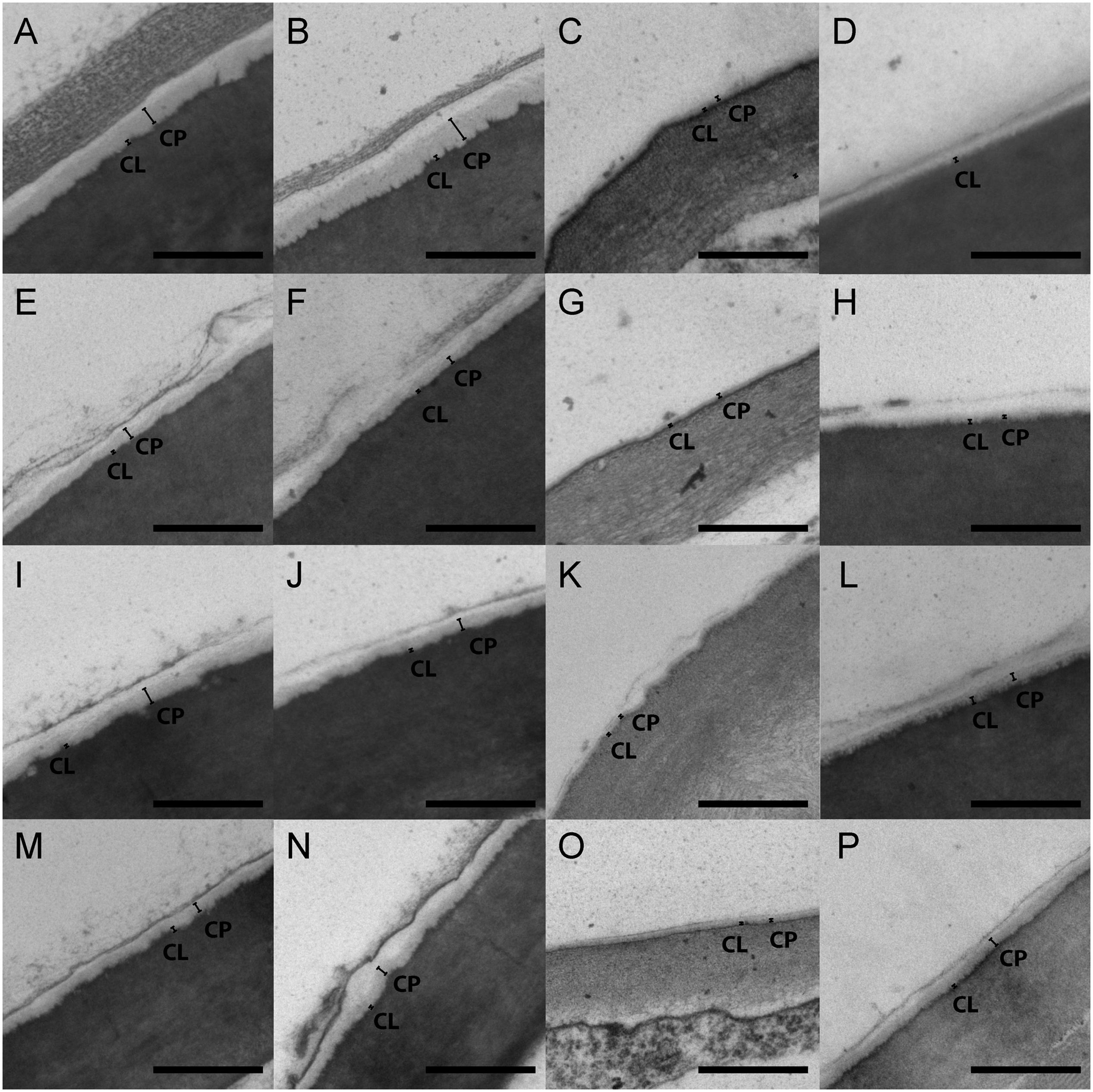
Figure two. Manual electron micrographs of moss cuticles. (A–D) Funaria hygrometrica. (Eastward–H) Physcomitrium pyriforme. (I–L) Physcomitrellopsis africana. (M–P) Aphanorrhegma serratum. (A, E, I, M) Young calyptra. (B, F, J, Northward) Older calyptra. (C, M, G, O) Immature sporophyte. (D, H, L, P) Older sporophyte. CL, cuticular layer; CP, cuticle proper. Calibration bars: 500 nm.
Morphology
Sporophyte sizes ranged from less than 1 mm tall in A. serratum to approximately 30 mm tall in F. hygrometrica (Tabular array 1; Figure 1). Taller sporophytes were associated with larger calyptra, with calyptra ranging from less than 0.5 mm in A. serratum to almost 4.v mm in F. hygrometrica (Table 1; Effigy 1).
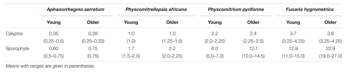
Table 1. Sizes in millimeters of calyptra and sporophytes at the 2 developmental stages analyzed in the study ( Northward = 5 for each).
Evolution
Comparisons of the cuticle layers of young and older calyptrae revealed no significant differences between the two developmental stages for the four species (Figure three). Thus, in all subsequent analyses data from the immature and older calyptra were combined. Meaning differences in the cuticle layers were institute between the young and older sporophytes for three of the four Funariaceae species (Effigy 3). Both cuticle layers of the older sporophytes had a larger average thickness compared to the young sporophytes for A. serratum and P. africana (Figures 3A,B), whereas the cuticle proper was significantly different only for P. pyriforme (Figure 3C). No significant differences in the jail cell lumen sizes or wall thicknesses were institute between immature and older calyptra, whereas lumen sizes and wall thicknesses were significantly increased between the young and older sporophytes (information not shown).
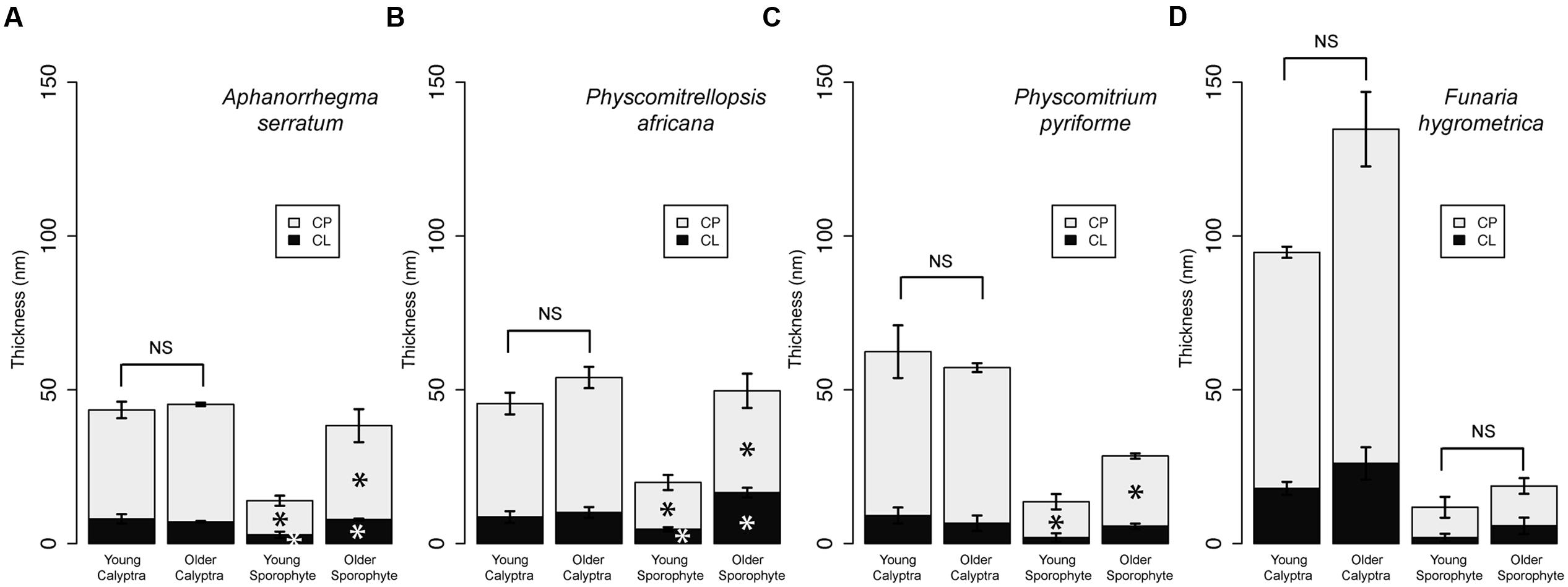
FIGURE iii. Cuticle thicknesses for calyptrae and sporophytes at ii developmental stages for four Funariaceae species. For each developmental stage (young and older), cuticle thicknesses were measured from the periclinal jail cell walls of the epidermis for three individuals and and then averaged (hateful ±SE). Statistically significant differences (P < 0.05) between cuticle layers are starred with an asterisk. (A) Aphanorrhegma serratum. (B) Physcomitrellopsis africana. (C) Physcomitrium pyriforme. (D) Funaria hygrometrica. CL, cuticular layer; CP, cuticle proper; NS, no pregnant differences.
Species Comparisons
Meaning differences between species characterize the calyptra cuticle (ANOVA: CP, F 3,twenty = 24.97, P < 0.001; CL, F 3,20 = 13.07, P < 0.001; full cuticle, F 3,20 = 23.12, P < 0.001; Figure 4A). Specifically the cuticle layers of F. hygrometrica were significantly thicker than the cuticles of the other three species (Effigy 4A).
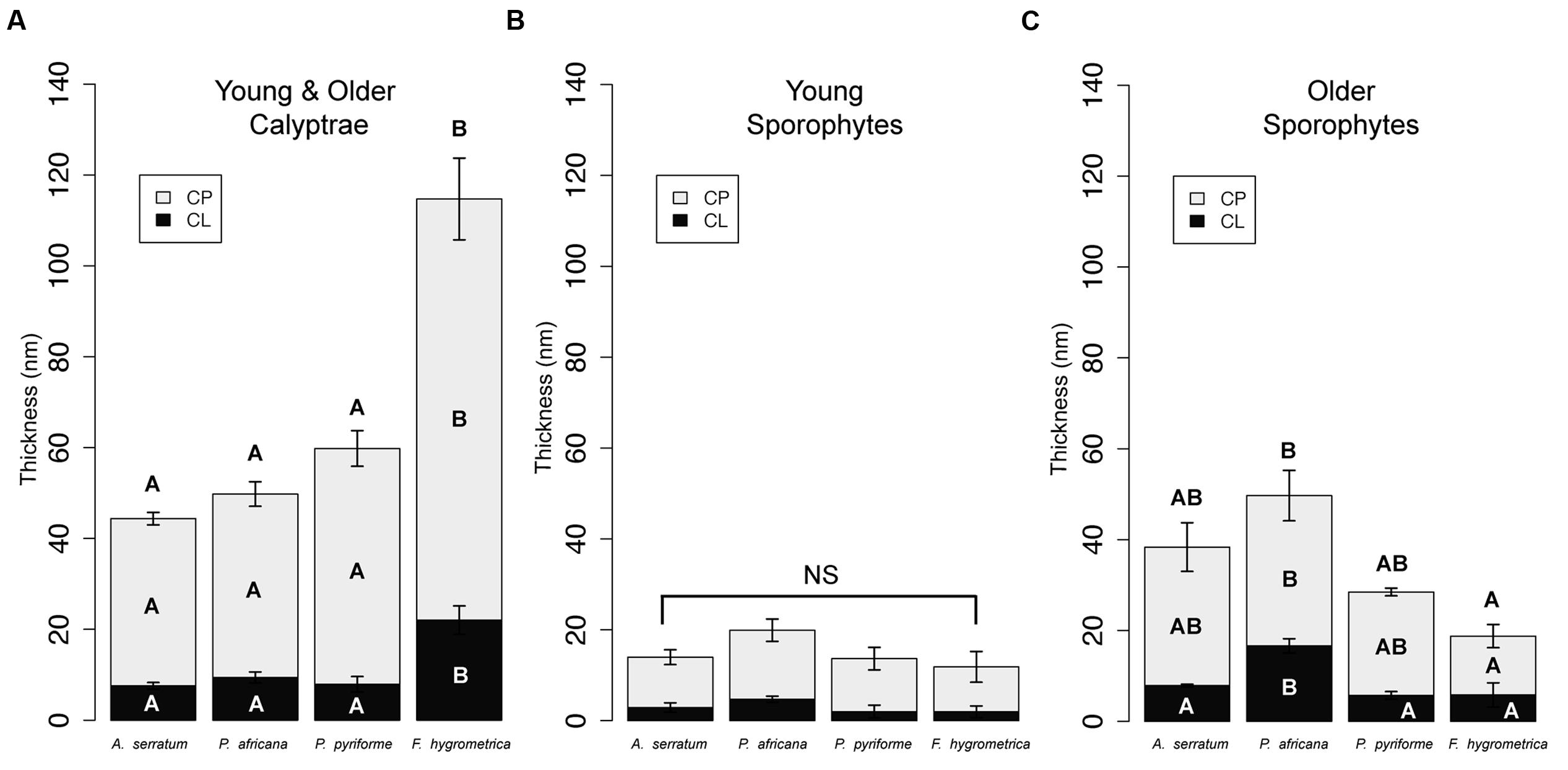
FIGURE 4. Cuticle thicknesses for calyptrae and sporophytes for 2 developmental stages for four Funariaceae species. (A) Calyptrae (young and older data combined). (B) Young sporophytes. (C) Older sporophytes. For each species, cuticle thicknesses were measured from the periclinal cell walls of the epidermis. Average cuticle thicknesses (mean ± SE) were calculated for 6 individuals per species for the calyptra (North = 24), combining data from young and older calyptrae, and three individuals per species for the young and older sporophytes (Northward = 24). CL, cuticular layer; CP, cuticle proper; NS, no significant differences; messages inside bars indicate layers that are significantly different based on Tukey HSD post-hoc tests (P < 0.05) for cuticle layers with significant ANOVAs.
Immature sporophyte cuticles were not significantly different across species (Figure 4B), whereas the cuticles of older sporophytes were significantly dissimilar (Figure 4C; ANOVA: CP, F 3,8 = 4.89, P < 0.05; CL, F three,viii = 10.23, P < 0.01; total cuticle, F iii,8 = 6.16, P < 0.05). The cuticle proper of P. africana was significantly thicker than F. hygrometrica, only the other two species were not significantly unlike from any other species. The cuticular layer was significantly thicker on P. africana compared to the other three species.
Sporophyte Height as a Proxy for Dehydration Stress
Sporophyte summit and calyptra length were significantly correlated at both developmental stages (Figure 5A; Young, adjusted R 2 = 0.98, df = 3, P < 0.01; Older, adjusted R two = 0.91, df = 3, P < 0.05). Cuticle thickness of the maternal calyptra was as well correlated with sporophyte pinnacle. Calyptra cuticle thickness data were combined from both developmental stages. The calyptra cuticle proper and cuticular layer thicknesses were significantly correlated with sporophyte height (Figures 5B,C; CP, adjusted R ii = 0.79, df = 23, P < 0.001; CL, adjusted R two = 0.50, df = 23, P < 0.001). No significant relationships were found between sporophyte height and sporophyte cuticle thickness at both the young and older developmental stages (Effigy 5D; CP, Young, adjusted R2 = –0.005, df = 11, P = 0.35; Older, adapted R 2 = 0.59, df = 11, P < 0.01; Figure 5E CL, Immature, adjusted R 2 = 0.05, df = 11, P = 0.23; Older, adjusted R 2 = 0.22, df = 11, P = 0.07).
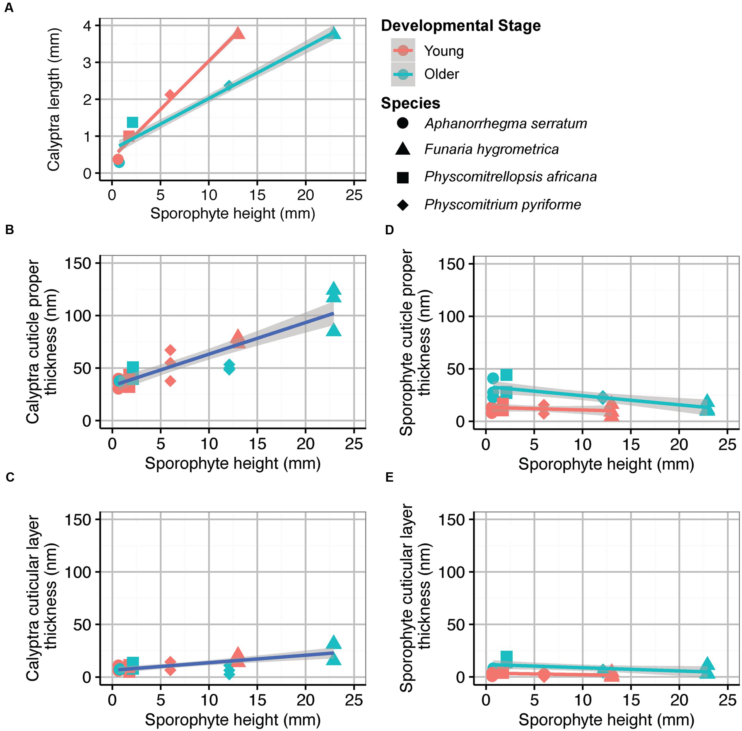
FIGURE 5. Average organ sizes and cuticle thicknesses for 4 Funariaceae species (circle Aphanorrhegma serratum , triangle Funaria hygrometrica , square Physcomitrellopsis africana , diamond Physcomitrium pyriforme ) at two developmental stages (young in red, older in greenish). Sporophyte height and calyptra length were averaged from v individuals of each species at each developmental stage (N = 40). Cuticle thicknesses were measured from the periclinal cell walls of the epidermis for three individuals per species at each developmental stage (N = 24). All regression lines are surrounded by 95% confidence intervals in gray (young in red, old in green, combined in blue). (A) Sporophyte height correlated with calyptra length; Young, adjusted R two = 0.98, df = 3, P < 0.01; Older, adjusted R 2 = 0.91, df = 3, P < 0.05. (B) Sporophyte height correlated with calyptra cuticle proper thickness; adapted R two = 0.79, df = 23, P < 0.001. (C) Sporophyte pinnacle correlated with calyptra cuticular layer thickness; adapted R two = 0.48, df = 22, P < 0.001. (D) Sporophyte height correlated with sporophyte cuticle proper thickness; Young, adjusted R 2 = -0.005, df = 11, P = 0.35; Older, adjusted R 2 = 0.59, df = eleven, P < 0.01. (Due east) Sporophyte height correlated with sporophyte cuticular layer thickness; Young, adjusted R 2 = 0.05, df = 11, P = 0.23; Older, adjusted R 2 = 0.22, df = xi, P = 0.07.
Discussion
The calyptra is a maternal organ covering the noon of the moss sporophyte, thereby protecting the young sporophyte offspring from aridity at least until meiosis occurs in the apical sporangium. This structure, which is derived from the archegonium and tissues of the leafy gametophyte below, is covered by a relatively thick cuticle (Budke et al., 2011), which develops early compared to the cuticle of the underlying sporophyte (Budke et al., 2012), and it is disquisitional for sporophyte fettle (Budke et al., 2013). These observations, based on F. hygrometrica, advise that the calyptra is under strong selection. Thus, variation in the calyptra cuticle may exist correlated with changes in sporophyte compages, such that species with short sporophytes that mature surrounded past the vegetative leaves and hence inside the laminar boundary layer, may be covered by a calyptra with a thinner cuticle and conversely that tall sporophytes elevating the sporangium higher up the laminar boundary layer prior to maturation may be protected by a thicker calyptra cuticle. We acknowledge that other aspects of the moss cuticle, including wax or cutin quantity and the ratios between waxes classes, may predict cuticular permeability (Riederer and Schreiber, 2001; Parsons et al., 2012). The present comparative study of four species provides the first insights into the relationship between cuticle thickness and sporophyte acme in mosses.
The observations past Budke et al. (2012) that young sporophytes accept relatively sparse cuticles that practise not thicken until late during capsule expansion and that the maternal gametophyte calyptra produces a mature cuticle early during sporophyte evolution that persists through sheathing expansion are confirmed for all taxa (Figure 3), regardless of sporophyte size. Taller sporophytes, which probable experience higher levels of dehydration stress (Niklas, 1994), have thicker calyptra cuticles (Figures 5B,C). The measurements of the epidermal cell sizes and cell wall thicknesses ostend our previous developmental observations (Budke et al., 2012); the calyptra is a static structure whose cells practise not significantly increase in size or develop thicker walls after detachment from the leafy gametophyte, whereas the sporophyte is undergoing dramatic changes during elongation and maturation in terms of epidermal cell size and wall thickness (information not shown). These observations are mirrored by our cuticle data, which show an early developing and and so static calyptra cuticle contrasting with a dynamic, late developing sporophyte cuticle (Figure 3). These observations from a broader taxon sampling further back up the hypothesis that the early maturing calyptra cuticle functions to protect the sporophyte apex from dehydration during early development, when moss sporophytes lack the structural protection provided by a thick cuticle (Budke et al., 2012, 2013).
Dispersing seeds and spores far away from the maternal constitute both decreases resource competition and aids in the colonization of novel locations (Janzen, 1970). One way to attain larger dispersal distances is by increasing plant tiptop (Thomson et al., 2011). In mosses, sporophyte summit varies both beyond (e.grand., Fife, 1982) and inside species (due east.thousand., Shaw, 1990). Even a small increment in sporophyte height may be enough to raise the spore filled capsule above the still air of the laminar boundary layer into more turbulent air flow, which would increment the ability of vertical updrafts to facilitate long distance dispersal events (Tackenberg, 2003). Increases in sporophyte height also increase the transpirational pull of resources from the maternal plant (Haig, 2012). Maximizing the resources an offspring acquires straight impacts sporophyte reproductive success. The potentially negative upshot is that taller sporophytes have increased exposures to dehydration stress. This is especially dangerous during the stage of stalk-building when the sensitive sporophyte apex is elevated across the protection of the laminar purlieus layer and the protective leaves of the maternal plant. At this stage protective structures are critical for the sporophyte to avert and ultimately survive the stress of dehydration. In mosses, taller species have both a thicker calyptra cuticle in addition to a larger calyptra (Figure 5). The ability to develop taller sporophytes likely shapes the efficiency of colonizing new habitats through effective spore dispersal, and thus may positively bear upon sporophyte reproductive success.
Tall sporophytes ascend from a prolonged period of seta development, ultimately delaying sheathing differentiation. During this phase, the presumptive sporangial tissues remain undifferentiated for a longer flow of time compared to taxa with short and more rapidly developing sporophytes. Taller sporophytes thus have a longer menstruum of vulnerability to damage and stress. A functional cuticle tin exist maintained longer either past repairing cuticle impairment (Hallam, 1970; Latimer and Severson, 1997; Neinhuis et al., 2001) or by initially producing a thicker cuticle that is more resistant to damage (Onoda et al., 2012). Overall, we did not find whatsoever significant increases or decreases in calyptra cuticle thickness across development (Figure 3). Though the calyptra can remain alive after detachment (Truthful, 1906; Bopp, 1954; Oehlkers and Bopp, 1957; Wynne and Budke, 2012), information technology ultimately dies and may lack the resources or ability to repair cuticle impairment. Alternatively, the calyptra cuticle is significantly thicker than the cuticle on all other parts of the maternal gametophyte (Budke et al., 2011; Buda et al., 2013) and we observed that species with taller sporophytes and thus longer periods of sporophyte development accept thicker calyptra cuticle layers (Figures 4A, 5). These observations marshal with a strategy of early investment in a thick calyptra cuticle that maintains its protective functions over longer periods of time by resisting damage.
Even within an individual, cuticle development is highly plastic and can be influenced by the surrounding environment (due east.yard., shade vs. sun leaves in Quercus, Osborn and Taylor, 1990; submerged vs. aeriform leaves in amphibious plants, Frost-Christensen et al., 2003). The leafy gametophytes of mosses can also increase their cuticle investment in response to alternating cycles of hydration and dehydration stress (Xu et al., 2009). The ability to alter cuticle development in response to stressful conditions could be advantageous for maternal moss plants, both improving their own reproductive success and the fettle of their offspring sporophyte. Our developmental observations reported here are from plants grown in common garden conditions, thus all differences in the cuticles can be attributed solely to taxon and tissue differences, not ecology influences. The influences that shape cuticle development on the maternal calyptra specifically and in bryophytes broadly are areas ripe for exploration. Expanding our knowledge of the environmental factors that impact cuticle development will enable us to better understand the physiology and evolution of protective strategies in plants.
In bryophytes, the sporophyte remains physically attached to the maternal gametophyte throughout its lifespan. Young sporophytes do photosynthesize; withal, they are dependent on nutrients and water from the maternal constitute (Ligrone and Gambardella, 1988). This presents a conflict over resources between the offspring and maternal plants (Haig and Wilczek, 2006; Haig, 2012), especially for species with perennial gametophytes that volition reproduce in subsequent years. The cuticle on the maternal calyptra may not simply play a protective role in aridity, but this layer may concurrently decrease sporophyte transpiration; reducing the resources taken by the offspring sporophyte from the maternal plant. On the opposite side of this conflict, the offspring potentially increases transpiration, and thus its pull of resources from the maternal plant, past increasing the number of stomata on the capsule or by increasing seta length, elevating the capsule farther in a higher place the boundary layer. Our data on the calyptra cuticle directly marshal with the predictions of this conflict. We observed that the calyptra cuticle is thicker for species with taller sporophytes, which may enable them to limit the transpirational pull of resources past the offspring from the maternal plant (Figure 4A).
The evolution of sporophyte morphology across the Funariaceae is widely thought to occur via the process of reduction (McDaniel et al., 2010; Liu et al., 2012; Beike et al., 2014; Medina et al., 2015). Parallel losses in structures that facilitate spore dispersal, such as peristome teeth, the operculum, and the seta, are observed across the family unit. The morphological reductions observed in both the maternal calyptra and offspring sporophyte of the Funariaceae could accept occurred nether several alternative scenarios; driven initially by morphological evolution of either the offspring sporophyte or the maternal calyptra or alternatively evolving in concert. In 1 scenario the evolution of a shorter sporophyte results in lower levels of dehydration stress, enabling the maternal gametophyte to invest fewer resource in the protective calyptra, by thinning the cuticle and ultimately developing a smaller calyptra. In an alternative scenario, the evolution of a smaller calyptra with a thinner calyptra cuticle results in higher levels of sporophyte dehydration stress, constraining and ultimately reducing sporophyte height. A well-resolved phylogeny combined with comparative methods may enable us to decide the most likely scenario (Felsenstein, 1985). In either case, the cuticle represents a costly structural investment, the lipids of which may crave more than than double the glucose for a found to build compared to cell wall polysaccharides (Poorter and Villar, 1997). Thus, any subtract in cuticle investment frees up resource to exist allocated to other developmental, reproductive, or physiological processes.
Many maternal organisms provide protection for their offspring and this study highlights a unique example of maternal protection in plants. The maternal calyptra is not a vestigial construction, simply has been retained and elaborated beyond the 12,500 species of mosses (Crosby et al., 1999). Using a comparative developmental framework we have expanded our knowledge of moss cuticle evolution to a broader number of taxa. This study lays the groundwork for future studies of morphologically and ecologically various species to ultimately further our agreement of the connections between maternal structures and their functional importance for offspring plants. Our observations augment our cognition of the plant cuticle and highlight the functionally important role the cuticle plays in preventing dehydration fifty-fifty in the relatively atomic bryophytes.
Author Contributions
JB designed, performed, and analyzed the experiments. JB and BG conceived the written report and wrote the manuscript. All authors read and approved the final version of the manuscript to exist published.
Funding
This enquiry was supported past a Katherine Esau Postdoctoral Fellowship to JB and a grant to BG (DEB-1146295) from the US National Science Foundation. Assistance from the faculty and staff of the University of Connecticut Electron Microscopy Facility was appreciated.
Conflict of Interest Statement
The authors declare that the inquiry was conducted in the absence of any commercial or financial relationships that could be construed as a potential disharmonize of involvement.
Acknowledgments
Nosotros give thanks Terry Hedderson and Nicholas Wilding for facilitating fieldwork in South Africa and enabling the collection of P. africana, and William Buck for providing a sample of A. serratum. We besides thank members of the Goffinet laboratory and reviewers for their helpful comments on before versions of this manuscript.
Footnotes
- ^http://rsb.info.nih.gov/ij/
References
Beike, A. K., von Stackelberg, M., Schallenberg-Rüdinger, Thou., Hanke, S. T., Follo, M., Quandt, D., et al. (2014). Molecular bear witness for convergent evolution and allopolyploid speciation within the Physcomitrium-Physcomitrella species complex. BMC Evol. Biol. 14:158. doi: x.1186/1471-2148-14-158
PubMed Abstract | CrossRef Full Text | Google Scholar
Bopp, Grand. (1954). Die wirkung von maleinhydrazid und kalyptraextrakt auf die verdickung von laubmoossporogonen. Naturwissenschaften 41, 243–245. doi: x.1007/BF00635130
CrossRef Total Text | Google Scholar
Buda, G. J., Barnes, W. J., Fich, Eastward. A., Park, S., Yeats, T. H., Zhao, Fifty., et al. (2013). An ATP bounden cassette transporter is required for cuticular wax deposition and desiccation tolerance in the moss Physcomitrella patens. Plant Cell 25, 4000–4013. doi: 10.1105/tpc.113.117648
PubMed Abstract | CrossRef Full Text | Google Scholar
Budke, J. M., Goffinet, B., and Jones, C. S. (2011). A hundred-year-old question: is the moss calyptra covered past a cuticle? A instance study of Funaria hygrometrica. Ann. Bot. 107, 1279–1286. doi: 10.1093/aob/mcr079
PubMed Abstract | CrossRef Full Text | Google Scholar
Budke, J. M., Goffinet, B., and Jones, C. S. (2012). The cuticle on the gametophyte calyptra matures before the sporophyte cuticle in the moss Funaria hygrometrica (Funariaceae). Am. J. Bot. 99, 14–22. doi: 10.3732/ajb.1100311
PubMed Abstruse | CrossRef Total Text | Google Scholar
Budke, J. M., Goffinet, B., and Jones, C. S. (2013). Dehydration protection provided by a maternal cuticle improves offspring fettle in the moss Funaria hygrometrica. Ann. Bot. 111, 781–789. doi: 10.1093/aob/mct033
PubMed Abstract | CrossRef Full Text | Google Scholar
Busta, L., Budke, J. M., and Jetter, R. (2016). Identification of β-hydroxy fatty acid esters and primary, secondary-alkanediol esters in cuticular waxes of the moss Funaria hygrometrica. Phytochemistry 121, 38–49. doi: 10.1016/j.phytochem.2015.10.007
PubMed Abstruse | CrossRef Full Text | Google Scholar
Campbell, C. L., Huang, J., and Payne, G. A. (1980). "Defence force at the perimeter: the outer walls and the gates," in Establish Illness: An Advanced Treatise: How Plants Defend Themselves, Vol. V, eds J. G. Horsfall and E. B. Cowling (New York, NY: Academic Press), 103–120.
Google Scholar
Crosby, Thou. R., Magill, R. E., Allen, B., and He, South. (1999). A Checklist of the Mosses. St. Louis, MO: Missouri Botanical Garden Press.
Google Scholar
Domínguez, E., Heredia-Guerrero, J. A., and Heredia, A. (2011). The biophysical design of plant cuticles: an overview. New Phytol. 189, 938–949. doi: ten.1111/j.1469-8137.2010.03553.x
PubMed Abstract | CrossRef Total Text | Google Scholar
Fife, A. J. (1982). A Generic Revision of the Funariaceae (Bryophyta: Musci). Ph.D. dissertation, University of Michigan, Ann Arbor, MI.
Google Scholar
French, J. C., and Paolillo, D. J. Jr (1975). On the role of the calyptra in permitting expansion of capsules in the moss Funaria. Bryologist 78, 438–446. doi: ten.2307/3242166
CrossRef Full Text | Google Scholar
Frost-Christensen, H., Jogensen, Fifty. B., and Floto, F. (2003). Species specificity of resistance to oxygen diffusion in sparse cuticular membranes from amphibious plants. Found Cell Environ. 26, 561–569. doi: 10.1046/j.1365-3040.2003.00986.x
CrossRef Full Text | Google Scholar
Goffinet, B., Budke, J. Thou., and Newman, L. C. (2011). Micromitriaceae: a new family of highly reduced mosses. Taxon 60, 1245–1254.
Google Scholar
Graham, 50. E. (1993). Origin of Land Plants. New York, NY: Wiley.
Google Scholar
Guzmán, P., Fernández, Five., Graça, J., Cabral, V., Kayali, North., Khayet, M., et al. (2014). Chemical and structural assay of Eucalyptus globulus and E. camaldulensis leaf cuticles: a lipidized cell wall region. Front. Found Sci. v:481. doi: 10.3389/fpls.2014.00481
PubMed Abstract | CrossRef Full Text | Google Scholar
Holmes, M. G., and Keiller, D. R. (2002). Effects of pubescence and waxes on the reflectance of leaves in the ultraviolet and photosynthetic wavebands: a comparison of a range of species. Plant Cell Environ. 25, 85–93. doi: x.1046/j.1365-3040.2002.00779.x
CrossRef Full Text | Google Scholar
Janzen, D. H. (1970). Herbivores and the number of tree species in tropical forests. Am. Nat. 104, 501–528. doi: 10.1086/282687
CrossRef Total Text | Google Scholar
Jeffree, C. East. (2006). "The fine structure of the plant cuticle," in Biology of the Plant Cuticle, Vol. 23, eds M. Riederer and C. Müller (Oxford: Blackwell Publishing), 11–125.
Google Scholar
Krauss, P., Markstädter, C., and Riederer, Thou. (1997). Attenuation of UV radiation past plant cuticles from woody species. Establish Cell Environ. 20, 1079–1085. doi: 10.1111/j.1365-3040.1997.tb00684.x
CrossRef Total Text | Google Scholar
Latimer, J. G., and Severson, R. F. (1997). Effect of mechanical and wet-stress workout of growth and cuticle limerick of broccoli transplants. J. Am. Soc. Hortic. 122, 788–791.
Google Scholar
Liu, Y., Budke, J. M., and Goffinet, B. (2012). Phylogenetic inference rejects sporophyte based nomenclature of the Funariaceae (Bryophyta): rapid radiation suggests rampant homoplasy in sporophyte evolution. Mol. Phylogenet. Evol. 62, 130–145. doi: 10.1016/j.ympev.2011.09.010
PubMed Abstruse | CrossRef Total Text | Google Scholar
McDaniel, Southward. F., von Stackelberg, K., Richardt, S., Quatrano, R. S., Reski, R., and Rensing, South. A. (2010). The speciation history of the Physcomitrium–Physcomitrella species complex. Evolution 64, 217–231. doi: x.1111/j.1558-5646.2009.00797.ten
PubMed Abstract | CrossRef Full Text | Google Scholar
Medina, R., Liu, Y., Li-Vocal, W., Shuiliang, K., Hylander, K., and Goffinet, B. (2015). Dna based revised geographic circumscription of species of Physcomitrella s.l. (Funariaceae): P. patens new to Eastern asia and P. magdalenae new to East Africa. Bryologist 118, 22–31. doi: 10.1639/0007-2745-118.1.022
CrossRef Full Text | Google Scholar
Niklas, G. (1994). Plant Allometry: The Scaling of form and Process. Chicago, IL: The University of Chicago Press.
Google Scholar
Oehlkers, F., and Bopp, Yard. (1957). Entwicklungsphysiologische untersuchungen an mossmutanten II. Die korrelation zwischen sporogon und kalyptra bei mutanten von Funaria und Physcomitrium. Z .Inductive Abstamm. Ver. 88, 608–618.
Onoda, Y., Richards, L., and Westoby, M. (2012). The importance of foliage cuticle for carbon economy and mechanical force. New Phytol. 196, 441–447. doi: 10.1111/j.1469-8137.2012.04263.10
PubMed Abstruse | CrossRef Total Text | Google Scholar
Osborn, J. Grand., and Taylor, T. (1990). Morphological and ultrastructural studies of institute cuticular membranes. I. Sunday and shade leaves of Quercus velutina (Fagaceae). Bot. Gaz. 151, 465–476. doi: x.1086/337846
CrossRef Total Text | Google Scholar
Parsons, E. P., Popopvsky, South., Lohrey, G. T., Lü, Southward., Alkalai-Tuvia, S., Perzelan, Y., et al. (2012). Fruit cuticle lipid limerick and fruit post-harvest water loss in an advanced backcross generation of pepper (Capsicum sp.). Physiol. Establish. 146, 15–25. doi: 10.1111/j.1399-3054.2012.01592.ten
PubMed Abstract | CrossRef Total Text | Google Scholar
Pfündel, E. E., Agati, M., and Cerovic, Z. G. (2006). "Optical Properties of Plant Surfaces," in Biological science of the Establish Cuticle, eds M. Riederer and C. Müller (Oxford: Blackwell Scientific), 216–249.
Google Scholar
Poorter, H., and Villar, R. (1997). "The fate of acquired carbon in plants: chemic composition and structure costs," in Establish Resources Allocation, eds F. A. Bazzaz and J. Grace (San Diego, CA: Academic Press),k39–72.
Google Scholar
Proctor, Thou. C. F. (1980). "Diffusion resistance in bryophytes," in Plants and Their Atmospheric Environs, eds J. Grace, Due east. D. Ford, and P. 1000. Jarvis (Oxford: Blackwell Scientific), 219–229.
Proctor, Yard. C. F. (1982). "Physiological ecology: h2o relations, light and temperature responses, carbon balance," in Bryophyte Ecology, ed. A. J. Eastward. Smith (London: Chapman and Hall), 333–382.
Google Scholar
R Development Cadre Team (2013). R: A Language and Surround for Statistical Computing, Version 3.1.2. Vienna: R Foundation for Statistical Computing.
Google Scholar
Renzaglia, K. Southward., McFarland, K. D., and Smith, D. Yard. (1997). Anatomy and ultrastructure of the sporophyte of Takakia ceratophylla (Bryophyta). Am. J. Bot. 84, 1337–1350. doi: 10.2307/2446132
PubMed Abstract | CrossRef Total Text | Google Scholar
Rice, S. Grand., and Schneider, North. (2004). Cushion size, surface roughness, and the control of water balance and carbon flux in the absorber moss Leucobryum glaucum (Leucobryaceae). Am. J. Bot. 91, 1164–1172. doi: 10.3732/ajb.91.8.1164
PubMed Abstract | CrossRef Total Text | Google Scholar
Riederer, Thousand., and Schreiber, L. (2001). Protecting confronting water loss: analysis of the bulwark properties of plant cuticles. J. Exp. Bot. 52, 2023–2032. doi: 10.1093/jexbot/52.363.2023
PubMed Abstruse | CrossRef Full Text | Google Scholar
Shaw, A. J. (1990). Intraclonal variation in morphology, growth charge per unit, and copper tolerance in the moss, Funaria hygrometrica. Evolution 44, 441–447. doi: 10.2307/2409421
CrossRef Full Text | Google Scholar
Smith Merrill, Thou. Fifty. (2007). "eight.2 Polytrichum," in Flora of North America, Bryophyta, Role i, Vol. 27, ed. Flora of N America Editorial Committee (New York, NY: Oxford University Press), 133–140.
Tackenberg, O. (2003). Modeling long-distance dispersal of plant diaspores past current of air. Ecol. Monogr. 73, 173–189. doi: 10.1890/0012-9615(2003)073[0173:MLDOPD]2.0.CO;2
CrossRef Full Text | Google Scholar
Thomas, R. J., Ryder, S. H., Gardner, M. I., Sheetz, J. P., and Nichipor, S. D. (1996). Photosynthetic office of leaf lamellae in Polytrichum commune. Bryologist 99, 6–xi. doi: x.2307/3244431
CrossRef Total Text | Google Scholar
Thomson, F. J., Moles, A. T., Auld, T. D., and Kingsford, R. T. (2011). Seed dispersal distance is more strongly correlated with found elevation than with seed mass. J. Ecol. 99, 1299–1307. doi: 10.1111/j.1365-2745.2011.01867.x
CrossRef Total Text | Google Scholar
True, R. H. (1906). Notes on the physiology of the sporophyte of Funaria and of Mnium. Beih. Bot. Centralbl. nineteen, 34–44.
PubMed Abstract | Google Scholar
Wynne, M. A., and Budke, J. K. (2012). Examining the power of calyptrae to produce protonema in Funaria hygrometrica. Evansia 29, 61–64. doi: 10.1639/079.029.0205
CrossRef Total Text | Google Scholar
Xu, S. J., Jiang, P. A., Wang, Z. W., and Wang, Y. (2009). Crystal structures and chemical limerick of leafage surface wax depositions on the desert moss Syntrichia caninervis. Biochem. Syst. Ecol. 37, 723–730. doi: 10.1016/j.bse.2009.12.012
CrossRef Full Text | Google Scholar

What Is A Sporophyte Apex,
Source: https://www.frontiersin.org/articles/10.3389/fpls.2016.00832/full
Posted by: wedelyoust1985.blogspot.com
0 Response to "What Is A Sporophyte Apex"
Post a Comment